Silica
coatings in the Ka’u Desert, Hawaii, a Mars analog terrain: a
micro-morphological, spectral, chemical and isotopic study
Steven M.
Chemtob, George R. Rossman, John M. Eiler
Division
of Geological and Planetary Sciences,
California Institute of Technology, Pasadena, CA
91125-2500,
U.S.A.
Bradley L. Jolliff, Raymond E. Arvidson
Washington University, Department of Earth and Planetary Sciences
St. Louis, MO, 63130, U.S.A.
Abstract
High-silica materials have been observed on
Mars, both from orbit by the CRISM spectrometer and in situ by the
Spirit rover at Gusev Crater. These observations potentially imply a
wet, geologically active Martian surface. To understand silica
formation on Mars, it is useful to study analogous terrestrial silica
deposits. We studied silica coatings that occur on the 1974 Kilauea
flow in the Ka’u Desert, Hawaii. These coatings are typically
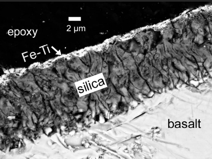
composed of two layers: a ~10 μm layer of amorphous silica,
capped by a ~1 μm layer of Fe-Ti oxide. The oxide coating is
composed of ~100 nm spherules, suggesting formation by chemical
deposition. Raman spectroscopy indicates altered silica glass as the
dominant phase in the silica coating, and anatase and rutile as
dominant phases in the Fe-Ti coating; jarosite also occurs within the
coatings. Oxygen isotopic contents of the coatings were determined by
secondary ion mass spectrometry (Cameca 7f and NanoSIMS). The measured
values, δ18OFe-Ti = 14.6±2.1‰, and δ18Osilica =
12.1±2.2‰ (relative to SMOW), are enriched in 18O
relative to the basalt substrate. The observations presented are
consistent with a residual formation mechanism for the silica coating.
Acid-sulfate solutions leached away divalent and trivalent cations,
leaving a silica-enriched layer behind. Micrometer-scale dissolution
and reprecipitation may have also occurred within the coatings.
Chemical similarities between the Hawaiian samples and the high-silica
deposits at Gusev suggest that the Martian deposits are the product of
extended periods of similar acid-sulfate leaching..
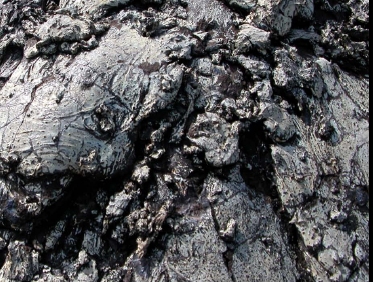
Coated basalt in the
field (left) and a SEM image of a cross section (right).


