
| |
|
|
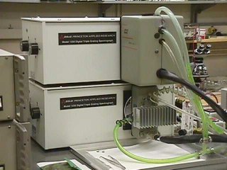 (Fig. 1) Diode Array Dectectors. 512 channel InGaAs on top, 1025 element Si on the bottom. |
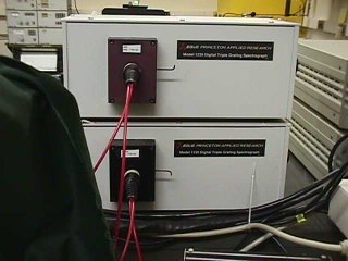 (Fig. 2) The spectographs. One for the Near-IR and one for the Visible-UV region. |
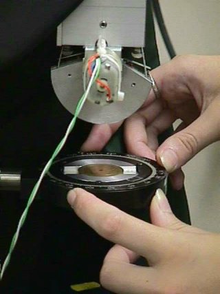 (Fig. 3) The sample location in the spectrometer. |
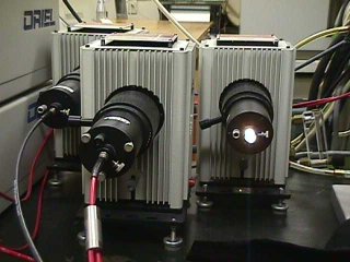 (Fig. 4) Light sources for the spectrometer system: Deuterium, Xeon Arc, and Tungsten lamps. |
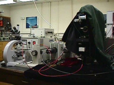 (Fig. 5) The whole system. |
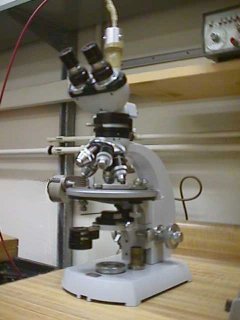 (Fig. 6) Optical microscope configured to work through fiber optics with the diode array spectrometer. |