George R. Rossman
Division of Geological and Planetary Sciences
California Institute of Technology
Pasadena, California, 91125, U.S.A.
ABSTRACT: Diopside with an unusual
violet color in hand specimen from south Baffin Island, Nunavut, Canada,
is found in calc-silicate lenses, associated with mirialite, pargasite,
phlogopite, calcite, apatite, titanite, talc, chlorite, plagioclase and
quartz. It occurs as massive aggregates of roughly equant grains. Basal
parting is evident and pyroxene cleavage is subtle. Indices of refraction
are na=1.670(1), nb=1.675(1),
and ng=1.695(1), 2VZ is
equal to 57.6(5)° at 589 nm. Pleochroism is weak to nonexistent. Dcalc
= 3.300 g/cm3. Cell dimensions determined from powder X-ray
diffraction are a = 9.730(4) Å, b = 8.873(3) Å, c = 5.275(2)
Å, b 109.95(3)°. A single-crystal
X-ray structural refinement was performed to determine bond lengths and
angles. The empirical formula, based on microprobe analysis and absorption
spectroscopy indicating 0.30% H2O in the structure, is (Ca0.96Na0.04)(Mg0.86Al0.06
Fe2+0.05Ti4+0.02) (Si1.89Al0.11)O5.93OH0.07.
The unusual colour is due to intervalence charge transfer between Fe2+
and Ti4+ at the M1 site, and is observed due to the low
overall concentration of Fe in the diopside. Cathodoluminescence indicates
that Mn2+ is present in the M1 site. Absorption spectroscopy
demonstrates that Mn3+ does not contribute to the violet colour.
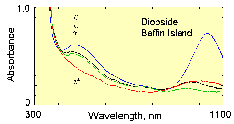
These spectra are taken in the directions of the alpha, beta and gamma indices of refraction: gamma-to-c = 42 degrees, beta = b-axis. Two slabs were used: (010) for E||a and E||g; and a slab perpendicular to c for the b = E||b-axis.
The violet color is due to the slightly higher transmission (less absorbance)
near 700 nm (red) than at 450 nm (blue). The band near 1050 nm in the b-direction
is due to Fe2+ in the M2 site. The band in a
near 2300 nm is also from Fe2+ in the M2 site. In the a
and g directions, there is a pair of M1-site
Fe2+ bands near 950 and 1100 nm.
In the a* direction (5K) (E vector vibrating perpendicular to both the b- and c-axes) there is essentially no aborption from the 480 nm band. This is an important point of comparison to the ADOR meteorite and speaks to the intervalence charge transfer. In this direction, there are not any adjacent cation sites, so IVCT can not occur. It should reach a maximum in the E||c direction if it occurs along the M1-M1 chains, and in the E||b direction if it is along the M1-M2 cation pairs.
Data Files as ASCII x,y data alpha 0.404 mm sample (77K file); beta 1.101 mm sample (79K file); gamma 0.404 mm sample (76K file); a* direction 1.101 mm (21K file).