Michail N. Taran* and George R. Rossman‡
*Institute of Geochemistry,
Mineralogy and Ore Formation
National Academy of Science of Ukraine
pr. Palladina 34
03142 Kyiv, Ukraine
‡Division of
Geological and Planetary Sciences
California Institute of Technology
Pasadena, CA 91125 USA
American Mineralogist 87, 1148-1153.
The Mineralogical Society of America
1015 18th St NW Ste 601, Washington, DC 20036-5274 USA
(http://www.minsocam.org)
The optical spectra of a dravite tourmaline
with an unusually high ratio of Fe3+ to Fe2+
and a synthetic elbaite
with high Fe3+ content were studied under high
temperature (297 to 600 K) and high pressure (to 10.51 GPa)
conditions. Individual absorption bands derived from paired iron
were identified in the spectra and their temperature and pressure
dependence studied. The most pronounced effects are the
intensification of the two bands derived from Fe2+
(~9090 cm-1 and ~14300 cm-1) at pressure,
their shifts to higher energy at pressure, and their pronounced
weakening at increasing temperature. Such behavior is assumed to
be caused by an electronic exchange interaction in a Fe2+-Fe3+
pair accommodated in adjacent Y sites of the structure.
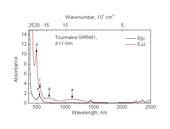
Temperature and pressure dependencies of the bands attributed to Fe3+-Fe3+ exchange-coupled pairs noticeably differ from those of Fe2+-Fe3+ pairs. This demonstrates that the two types of the pairs have different types of exchange interactions and points to the need for further experimental and theoretical investigations.
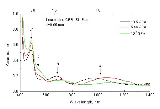
The intensity of the intense E^c-polarized band at ~ 20580 cm-1 originating from the 6A1g -> (4A1g, 4Eg) transition of the Fe3+(Y) -Fe3+(Y) pair, moderately depends on temperature and pressure. The intensity of a weak shoulder at ~18350 cm-1 (E^c), also attributed to a Fe3+(Y) -Fe3+(Y) pair, decreases and nearly disappears between 5.44 and 9.55 GPa. The intensity of E||c-polarized band at ~18500 cm-1, attributed to Fe3+(Z) -Fe3+(Z) pair, displays a strong inverse temperature dependence, whereas the energy of the band remains nearly constant.
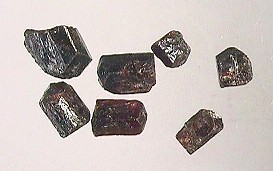
The dravite is from the the vicinity of Osarara, Narok
District, Kenya. The sample is from the US National Museum of
Natural History, Smithsonian Institution, sample number
126030.
Dravite, ideally, is NaMg3Al6(BO3)
3Si6O18(O,OH)4. This
crystal contains a major amount of Fe3+ substitution
and has a formula (Na0.81Ca0.01K0.01[]0.16)
Mg1.3Al0.95Fe3+0.56
Fe2+0.05Ti0.03Cr0.01
{Al5.09Mg0.91} (BO3)3
Si6 O18 (O,OH)4 according to
Hawthorne et al. (1993).
The optical
absorption spectrum of this dravite shows features
(absorption bands) that arise from an interaction between of
pairs of Fe3+ ions. These features are about 100 times
more intense than the features would be if the iron were not
paired with an adjacent ion.
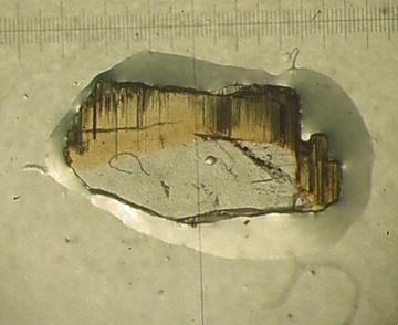
At 9.55 GPa, the color changes from orange to
greenish-grey.
 |
 |
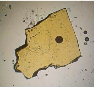
| At ambient temperature and pressure, the Kenyian dravite is orangish red in a 0.050 mm thick section oriented perpendicular to c. | The picture shows the sample at 9.55 GPa and a ruby chip (for pressure determination within a gasket hole. The black solid circle is the upper diaphragm of the microspectrometer (the measured part of the sample). |
As should be expected from the color change, the optical absorption spectrum shows that, at pressure, many of the bands shift energy and change in intensity.
References:
Bank H (1974) Rote Turmaline mit hoher Licht- und Doppelbrechung aus Kenya. Z. Dt. Gemmol. Gesell. 23, 89-92.
Dunn PJ, Arem JE, Saul J (1975) Red dravite from Kenya. J. Gemmology 14, 386-387.
Hawthorne FC, MacDonald DJ, Burns PC (1993) Reassignment of cation site occupancies in tourmaline: Al-Mg disorder in the crystal structure of dravite. American Mineralogist 78, 265-270.
Mattson SM and Rossman GR (1984) Ferric iron in tourmaline. Physics and Chemistry of Minerals 11, 225-234.